

Obstetrics scan( which is the main reason for the invention of ultrasound before we started using it to do other thing) the goal
is to see the both the foetus and the mother in good condition, hence the reason for first, second and third trimester scan before
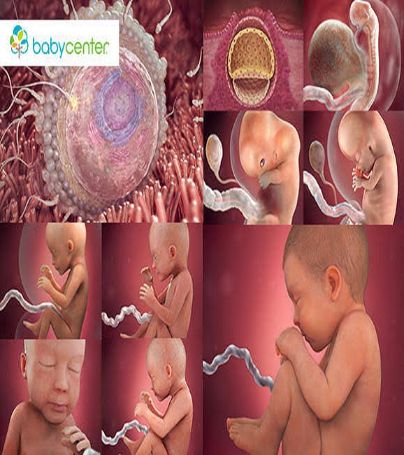
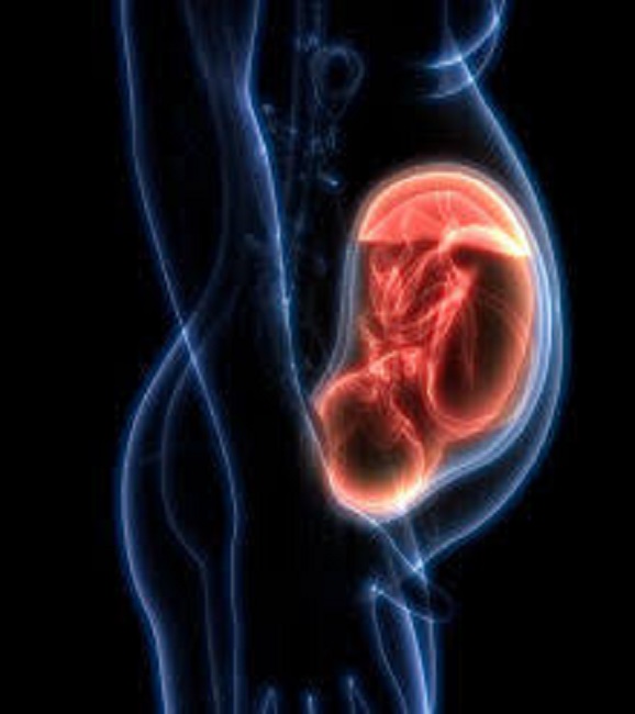
delivery. Most of us know where embryos begin their life. They start in the mother's womb. After spending around nine months, inside
her, the foetus is ready for the outside world. During one day our blood will make over 600 complete trips through the body.
But what happens in the mother to make the child begin? It is an interesting story of God and nature creating life. God fata heart rate
means good health.
Two tiny parts are needed to make a baby. They are too small to be seen well without a microscope. One part is called
the sperm cell. It comes from the father. The second part is the egg cell. It is made by the mother's body. Children are made
both the father and mother.
If we could see a sperm cell, it would look like a tiny tadpole. It has a small tail and swims quickly.
The sperm wants to find an egg and begin a baby. However, of the billions of sperm not many match up with the egg.
When these two cells come together, they fertilize. This means the tiny parts have started to make life. The two cells grows by
splitting and still staying together. the two become four, the four becomes sixteen.
They keep splitting for eight days and yet it is
only a small speck. You could put fifty of these on one inch of a ruler. When it is this size, the cells rest against the mother's womb.
There it will grow and live until it is time to be born.
The sperm and the egg are good for only 48 hours. If they do not meet and begin within two days. they die and new ones are needed.
The minutes these two cells meet most of the child's looks are put together. It hair, colour, sex, face and more are all settled.
As the cells grow they form three layers. The outside layer forms the skin. The middle layer makes the skeleton and nervous system.
The third layer will create our digestive tract.
Scientist have studied the making of a body and know a lot about it and a Sonographer is to use this knowledge to help the woman
in cyesis. In general examination, the investigations required include: abdominal, obstetric(routine), pelvic, renal, abdomino-pelvic,
scrotal, special e.g(gender differentiation carotid, thyroid, orbital and interventional scan e.g effusion or drainage. While others
include: droppler scan, colour flow scan and obstetric scan (comlications) and mamo scan.